


Current issue
Dental Press Endodontics – ISSN 2178-3713
Dental Press Endod.
v. 02, no. 4
October / November / September
2012
Editorial
The modern endodontic therapy
3 3
With the avalanche of new concepts, techniques and materials, the modern endodontic therapy — increasingly modern — suggests a single path to be followed so that one can achieve bigger recognition and distinction in the dental field: Professional updating. To be considered competent, a professional must have both scientific knowledge and technical skills; but it is not enough to give attention to the disease only, it is necessary to understand the patient as a whole. A dignified Endodontics practice involves taking positions and attitudes.
This professional updating occurs by reading periodicals, which aims at preserving science history, pointing results of scientific research and disseminating innovative materials and techniques to allow its clinical applicability. However, certain demands force the authors to publish quality papers in international journals, a fact contested by professionals, considering that the disseminators of knowledge must also have credibility in national periodicals, benefiting the endodontic class. In Brazilian Endodontics, this vehicle is the Dental Press Endodontics journal, which aims to be the responsible for knowledge transfer. Given the difficulties of publishing abroad, it is necessary that our researchers focus on the internal market and direct many of their studies to here, since it is the Brazilian endodontic professionals that subscribe to this dissemination vehicle and this is their demand.
The other way of being updated is to take part in events specific to the Endodontics area. Among these, the NATIONAL CIRCUIT OF ENDODONTICS aims to discuss current issues, important to the clinical practice of Endodontics, with renowned professors. In 2013, in its 5th edition, the event will be in Curitiba/PR - Brazil, from 25th to 27th of April. Seeking for clinical solutions, “The reason why I changed the technique” promises to show that the best option not always is the one proposed by the manufacturer. “Treatment of endodontic infection” with Profs. Helio Pereira Lopes and José Siqueira Jr. will be the main attraction. But “Why failures happen and how to solve them” promises a great debate in the search for solutions and on the various factors involved. We have no doubt that we will witness and take part in important moments for our professional growth. In today’s world, the scientific improvement is more than a fad, it is a must for professional’s survival.
Done. Now you have in your hands the two most important vehicles for your updating. Your patient deserves it!!
Endo in Endo
The waiting time for inducing orthodontic movement after endodontic treatment, even with perforations
Endodontics-Orthodontics. Orthodontic movement. Periapical repair. Endodontic repair.
11 14
The delay period required to start orthodontic movement after endodontic therapy has always raised many questions. This study aimed to reduce these questions, add some considerations into these discussions and suggest a delay period that agrees with the periodontal tissues biology. When the main goals of endodontic treatment are reached, regardless if the tooth presents pulp vitality, pulp necrosis, chronic root lesions or root perforations; 30 days after endodontic treatment, the periodontal tissue is in final repair stages, reorganizing the tissue to restore its physiology and anatomy, even though this process does not occur in such manner, since mineralization is incipient. Orthodontic forces should not biologically interfere with tissue repair, with the pathogenic and virulent microbes involved in pulp necrosis, with chronic periapical lesions and with perforated roots due to endodontic treatment.
Original articles
Effectiveness of 2% chlorhexidine gel compared to two solvents commonly used in endodontic retreatment
Retreatment. Chlorhexidine. Solvents. Root canal preparation.
15 19
Objective: The present study has compared the action of 2% chlorhexidine gel to two solvents commonly used in endodontic retreatment, xylol and eucalyptol, regarding the cleaning of the root canal walls.
Methods: Forty-five human single-rooted teeth were randomly divided into three groups. The teeth were instrumented, filled and radiographed before being stored in an oven at 37°C for 60 days. The filling material was removed from root canals according to the following techniques: Kerr and Hedströem files with xylol (G1), eucalyptol (G2), and 2% chlorhexidine gel (G3). After removing the filling material, the teeth were radiographed in ortho- and mesio-radial directions. The radiographs were analyzed by three double-blinded examiners for the presence of remaining filling material. Next, the teeth were longitudinally fractured and the resulting faces were digitally scanned. The Image Tool software was used to assess the amount of remaining filling material, and data were statistically analyzed.
Results: With regard to the radiographic analysis, no statistically significant differences were observed between the groups studied (i.e. chlorhexidine, xylol and eucalyptol). However, when digitalized images were analyzed, xylol was found to be significantly more efficient for cleaning the root canals compared to 2% chlorhexidine gel.
Conclusion: It can be stated that xylol was the most effective solution for removal of filing material compared to 2% chlorhexidine gel and eucalyptol.
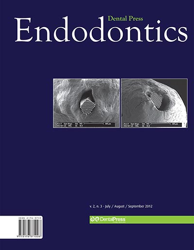
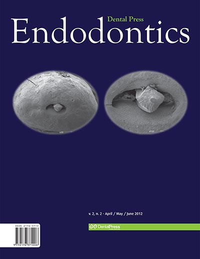
In vitro evaluation of dentin marginal adaptation of three root-end filling materials inserted with and without surgical microscope
Retrograde obturation. Scanning electron microscopy (SEM). Endodontics. Oral surgery.
20 25
Objective: Periradicular surgery is the procedure of choice when one aims to solve problems or complications which conventional endodontic therapeutics has not been able to solve. Surgical therapy comprises a number of procedures, among which retrofilling. The aim of this study was evaluating the degree of dentin marginal adaptation of root-end filling materials, as well as ascertaining the effectiveness of optical microscopic usage in the insertion of these materials.
Methods: Sixty upper canine teeth were selected, apicectomized and then rood-end cavities were prepared with the help of ultrasonic tips. The specimens were divided according to the material used: White MTA Angelus®, Super-EBA® and Sealapex® + White MTA Angelus®, it being that optical microscope was used in half of the samples of each group.
All samples were processed and evaluated by scanning electronic microscopy (SEM).
Results: The three materials tested presented satisfactory marginal adaptation. The use, or not, of the optical microscope, did not change the degree of adaptation of root-end filling materials evaluated in the present study.
Conclusion: All materials tested (White MTA Angelus®, Super-EBA®, and Sealapex® cement added to White MTA Angelus®) proved efficient regarding the issue evaluated, dentin marginal adaptation. The use of the optical facilitated insertion of root-end filling materials, due to better illumination and magnification. However, it did not promote any difference in the materials marginal adaptation to root-end cavities, when compared with its non-utilization.
Eugenol influence on the bond strength of intracanal metallic cast posts bonded with resinous cement
Dental materials. Endodontics. Intracanal posts. Cementation and bond strength.
26 31
Objective: To verify the influence of the eugenol on the bond strength of cast intracanal posts using resinous cement.
Methods: Root canal shaping of 33 human maxillary central incisors with 15 mm was performed standardizing the apical shaping at #55 file, 1 mm below de apical foramen. The teeth were divided in 3 experimental groups and 1 control. Group I (Control group) was composed by 3 teeth with root canal filling. The experimental groups were composed by 10 teeth each, filled by gutta-percha associated to 3 types of root canal sealers, used according to the group: Group II – epoxy resin based root canal sealer (AH Plus); Group III – calcium hydroxide based root canal sealer (Sealapex); Group IV – zinc oxide and eugenol based root canal sealer (Endofill). After the root canal preparation, 10 mm for the intracanal post, and the cast posts were adjusted and cemented with resinous cement (RelyX ARC). Every specimens were submitted to the mechanical test in the Universal Testing Machine Kratos 5002, at 0,5 mm/min speed and the values of the higher strength needed to dislocate the posts were registered and submitted to statistical analysis by the tests ANOVA and Tukey with 5% significance level.
Results: The control group presented mean of 598.05 kgf/cm2, AH Plus 475,43 kgf/cm2, Sealapex 358,03 kgf/cm2 and Endofill 213,70 kgf/cm2.
Conclusion: The eugenol influenced the bond strength of intracanal cast posts using resinous cement decreasing tensile resistance.
Efficiency of different concentrations of sodium hypochlorite during endodontic treatment. Literature review
Sodium hypochlorite. Antimicrobial activity. Organic solvent. Irrigation solutions.
32 37
The aim of this study is to evaluate, through a literature review, the effectiveness of various concentrations of sodium hypochlorite during endodontic treatment. It was possible to verify that the 0.5% sodium hypochlorite concentration needs more time to dissolve organic tissue while causing less irritation to periapical tissues. The 1% concentration showed lower loss of chlorine due to the presence of stabilizer, making the solution more reliable for long periods after open. The 2.5% concentration showed better bactericidal action and a good tissue dissolution time; the 5.25% concentration showed higher solvent potential and bactericidal effect, with lower surface tension and consequently better root canal decontamination. However, the highest concentration was also more toxic to periapical tissues, promoting greater irritation. Based on the literature review it can be said that the 2.5% sodium hypochlorite concentration, due to its less cytotoxic properties, is the most suitable for endodontic treatment of root canals.
Dental fracture stabilization for insertion of fiber-reinforced post and tooth-fragment reattachment: 6-month follow-up
Fracture. Reattachment. Adhesion. Glass fiber post
38 45
Dental fractures in endodontically treated teeth with a great loss of dental structure may be restored using bonding techniques associated with glass fiber post retention of the fragment. The present study reports a weakened and fractured crown restored by dental reattachment after crown stabilization with polyvinilsiloxane matrix, and a glass fiber post cementation for dental fragment retention. The employed techniques enabled the correct dental positioning during reattachment, with a possible increase of resistance. After 6 months follow-up the periodontal tissues showed itself in health condition and functional evaluations maintained the success of the proposed treatment.
Removal of a silver cone by using clinical microscope and ultrasound: Case report
Retreatment. Ultrasound. Microscopy.
46 50
Introduction: The retreatment of teeth with endodontic failure associated with the use of silver cone as filling material is still today a reality in the endodontic practice. The present work reports a case of endodontic failure resulting from the use of silver cone and subsequent endodontic retreatment.
Case report: The procedure consisted of removing the existing metal-ceramic crown and endodontic retreatment with removal of the silver cone, which was apically sectioned by using ultrasound and clinical microscope. Next, the root canal system was filled and glass-fiber posts and metal-ceramic crown were placed.
Conclusion: The use of microscope in association with ultrasound was crucial for performing the retreatment, thus allowing the silver cone to be safely removed without unnecessary wear of the dentinal structure.
The use of white MTA in the treatment of internal root resorption: Case report
Root resorption. Root canal obturation. Esthetics.
51 56
Introduction: Internal root resorption is a rare occurrence, asymptomatic, with slow progression, detected through routine radiographic examination, in which appears as a radiolucent and uniform lesion. The etiology and pathogenesis are not well understood, and it can occur as a result of trauma, orthodontic force, excess of heat, and other iatrogenic causes. After diagnosis of internal resorption, endodontic treatment is the choice.
Objective: This article aims to report the clinical case of a female patient, 19 years of age, underwent orthodontic treatment. After three years of treatment in the clinical and radiographic examination was verified the presence of internal root resorption in the tooth #22, which was asymptomatic.
Methods: Pulpectomy and changes of calcium hydroxide curative was performed. After six months of the beginning of treatment, with the tooth asymptomatic, radiographically stable and no bleeding, was performed the fill of the pulp cavity with white MTA. In esthetic recovery of the element, was chosen subepithelial connective tissue graft through the technique of plastic and microsurgery, subsequently a direct facet of composite resin was made.
Conclusion: Therefore, it can be concluded that the clinical and radiographic control of patients undergoing orthodontic treatment is important in the diagnosis of internal resorption. The White MTA presented itself as an excellent alternative for treatment of internal resorption, assisted with esthetic treatments.
Treatment of a lately replanted avulsed permanent tooth: Case report
Dental avulsion. Dental replantation. Facial traumas.
57 64
Avulsion after dental trauma is one of the most serious emergencies in the dental offices. Treatment success for this kind of injury lies on explaining to patients and dentists about the prompt management of the avulsed tooth. In the present study it’s emphasized the relationship between the whishes of the patient and his parents and the maintenance of the avulsed tooth in the oral cavity, exerting its esthetic and masticatory functions. Besides the time, other factors such as handling the tooth to be reimplanted, replantation technique, storage medium, endodontic treatment of the reimplanted tooth, intracanal medication and postoperative control contribute to a better prognosis. Dental avulsion usually affects children during their periods of bone growth, thus replantation aims to eliminate esthetic, psychological and social consequences for the patient, and can avoid prosthesis, satisfying patient’s whishes.
Healing of an extensive periapical lesion by means of conventional endodontic treatment
Calcium hydroxide. Radicular cyst. Root canal treatment.
65 69
Objective: The radicular cysts, also called peripheral cysts, are inflammatory cysts of the jaws, formed in the dental apices, with necrotic and infected pulps. The aim of this study was to report a clinical case of an extensive periapical lesion of endodontic origin, suggestive of peripheral cyst between teeth #33 and #34, near the region of the mental foramen.
Methods: The necropulpectomy endodontic treatment was performed no tooth #34, the dressing with calcium hydroxide paste was renewed on a monthly basis for 10 months, followed by endodontic obturation.
Results: After one year of conventional endodontic treatment the patient is without signs of recurrence.
Conclusions: The treatment endodontic provided a favorable clinical and radiographic response, without pain symptoms with the signs of regression of lesions, no need for additional surgery in the periapical region.